Macular degeneration 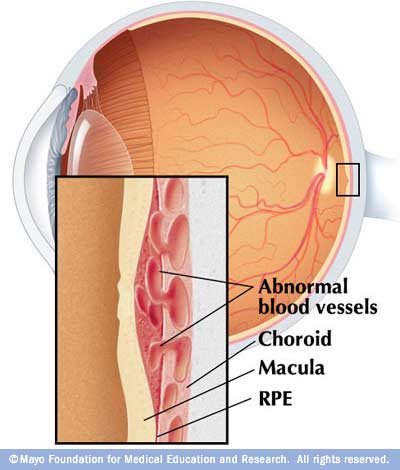
Wet macular degeneration
Associated with this condition in general with age, which usually affects older people, although some form Yaf'i them.
Established macular degeneration as a result of degradation of cones sensitive to light are located in the courtyard areata of the retina and Patch macula is the location of vision on the retina most sensitive to the details and colors, and macular degeneration there was a gradual details of the vision and the area of central vision (as opposed to Blue, where they inspected the edges external field of vision).
It affects both eyes, but one eye is usually affected by a few weeks before the other.
Signs and symptoms:
• The difficulty to focus on the written text.
• inability to recognize faces easily.
• Problems in identifying details when you watch TV.
Located between the eye and the placenta Cbugetha insulating layer and protective, with age, show some flaws on this layer, which allows some fluid to slip the eye retina itself. And this can cause the liquid to later damage and aggravated Balasyat sensitive cones, which is the primary key to generate a crisp, clear picture - a disorder known as Baltnks degeneration.
What is the spot?
Spot is an area in the center of the retina is very small, but very sensitive. Restricted to function specifically, to allow Ptbier objects in the center of your field of vision inside the eye.
Macular degeneration and leads to the increasing loss of sight in the central areas, but the outer edges of the field of vision remains relatively unaffected.
Discovery Status:
It is not always easy to identify the signs of macular degeneration, because the deterioration can happen very slowly and gradually over several years. But if you notice a gap or buckling in the central square of the vision, you should immediately rush to visit the doctor.
What can I do doctor?
Can a doctor or eye specialist to determine changes in the spot on the retina when the eyes examined Iwastp lens of the eye.
In some cases, be some other tests to assess the extent of damage to include a procedure known as angiography, fluorescein angiography Filorsin material where it is to take pictures of blood vessels in the eye.
And can be useful to laser treatment of the eye in some cases, even though he can stop the rampant disorder only, and not reverse the damage that may have occurred already.
Unfortunately, this situation is curable when most people.
Definitions
The structure of the retina:
The main theme of one layer of the eye sensitive to light located in the back of the eye called the retina retina. Network is composed of specialized cells called rods and cones generate electrical signals when exposed to light. Signals to be transmitted later on the length of the optic nerve to the brain, where it is processed.
Bacilli:
A light-sensitive cells, and share many of which connect one to the brain. These bacilli in dim light conditions, and leave information Black and white only, which is sensitive to movement, but do not give a very clear picture.
They are concentrated at the edges of network, hence the ability to see through the corners of the eye movements.
Cones:
Which cells respond to the colors - red, blue, and green. Each cell of Tusiltha Special to the brain, generating color images and clear, but it does not work well only in high optical wrenches.
Gather cones in the retina, and in particular at the point where the lens focalization image - known as the spot
0 commentaires